What Is Bursitis In The Foot?
Overview
Achilles tendon bursitis, or retrocalcaneal bursitis, can affect anyone, but is typically a foot condition in athletes, especially runners. Because of similar symptoms, this condition is often confused with Achilles tendinitis. At times, bursitis of the Achilles tendon can occur in conjunction with Achilles tendinitis. When both conditions are present, it is referred to as Haglund's syndrome. If you are a runner and are experiencing pain in your lower leg and heel area, you may be suffering from Achilles tendon bursitis. Proper treatment of the condition can help relieve the pain and allow your leg to heal.
Causes
Inflammation of the calcaneal bursae is most commonly caused by repetitive overuse and cumulative trauma, as seen in runners wearing tight-fitting shoes. Such bursitis may also be associated with conditions such as gout, rheumatoid arthritis, and seronegative spondyloarthropathies. In some cases, subtendinous calcaneal bursitis is caused by bursal impingement between the Achilles tendon and an excessively prominent posterior superior aspect of a calcaneus that has been affected by Haglund deformity.
Symptoms
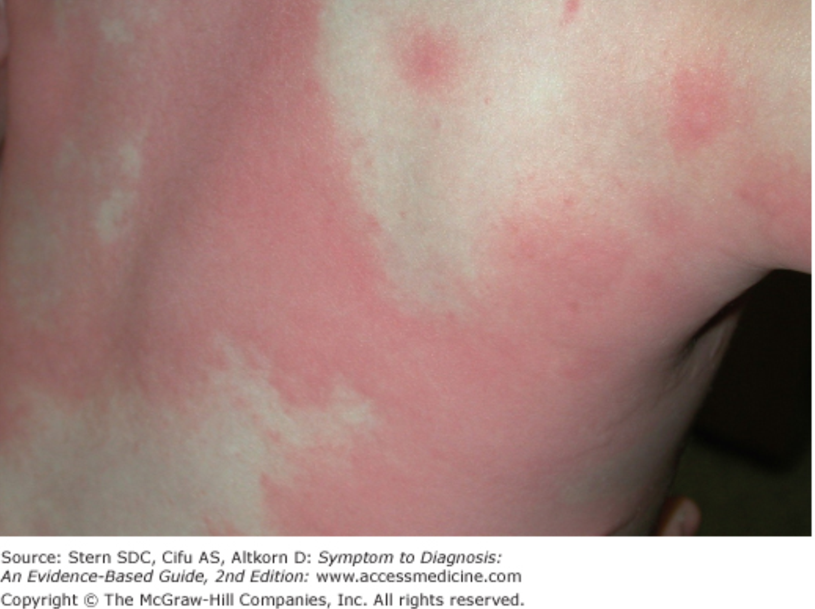
Bursitis usually causes a dull pain, tenderness, and stiffness near the affected bursa. The bursa may swell and make the skin around it red and warm to the touch. Bursitis is most common in the shoulder camera.gif, elbow camera.gif, hip camera.gif, and knee camera.gif. Bursitis may also occur near the Achilles tendon or in the foot. Symptoms of bursitis may be like those of tendinopathy. Both occur in the tissues in and around the joints. Check with your doctor if your pain is severe, if the sore area becomes very hot or red, or if you have a fever.
Diagnosis
In addition to a complete medical history and physical examination, diagnostic procedures for bursitis may include the following. X-ray. A diagnostic test that uses invisible electromagnetic energy beams to produce images of internal tissues, bones, and organs onto film. Magnetic resonance imaging (MRI). A diagnostic procedure that uses a combination of large magnets, radiofrequencies, and a computer to produce detailed images of organs and structures within the body. Ultrasound. A diagnostic technique that uses high-frequency sound waves to create an image of the internal organs. Aspiration. A procedure that involves removal of fluid from the swollen bursa to exclude infection or gout as causes of bursitis. Blood tests. Lab tests that are done to confirm or eliminate other conditions.
Non Surgical Treatment
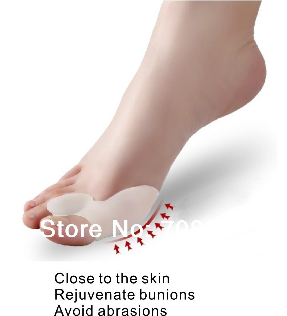
There are a variety of treatments for bursitis of the heel. Bursitis on the bottom of your heel (which is called infracalcaneal bursitis) is common in heels with thinning fat pads. Gel heel cushions or custom made orthotics (that have a horse-shoe cut and extra foam in the heel) can be lifesavers in reducing the pain. For bursitis of the posterior heel (retrocalcaneal bursitis), try to avoid going barefoot and to reduce the stress on the Achilles tendon by not over flexing your heel, the tighter your Achilles becomes, the more you compress the bursa sacs of the posterior heel. Heel lifts can help this, or wearing shoes with elevated heels (note that this method is not sanctioning high heels, as high heels can provide little comfort or support and usually are tight in the areas where your bursitis is most inflamed). Products such as AirHeel made by Aircast can help massage the bottom and back of the heel, helping to decrease pain.
Surgical Treatment
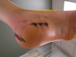
Only if non-surgical attempts at treatment fail, will it make sense to consider surgery. Surgery for retrocalcanel bursitis can include many different procedures. Some of these include removal of the bursa, removing any excess bone at the back of the heel (calcaneal exostectomy), and occasionally detachment and re-attachment of the Achilles tendon. If the foot structure and shape of the heel bone is a primary cause of the bursitis, surgery to re-align the heel bone (calcaneal osteotomy) may be considered. Regardless of which exact surgery is planned, the goal is always to decrease pain and correct the deformity. The idea is to get you back to the activities that you really enjoy. Your foot and ankle surgeon will determine the exact surgical procedure that is most likely to correct the problem in your case. But if you have to have surgery, you can work together to develop a plan that will help assure success.
Prevention
People can lower the risk of bursitis by gradually strengthening and stretching the muscles around the joints and taking regular breaks from repetitive motion that might irritate bursae. Prolonged time resting on the elbows or kneeling should be avoided, if it cannot be avoided, wearing cushioned elbow and knee pads can help protect the bursae. Comfortable, supportive, low-heeled shoes can help prevent bursitis in the foot.
Achilles tendon bursitis, or retrocalcaneal bursitis, can affect anyone, but is typically a foot condition in athletes, especially runners. Because of similar symptoms, this condition is often confused with Achilles tendinitis. At times, bursitis of the Achilles tendon can occur in conjunction with Achilles tendinitis. When both conditions are present, it is referred to as Haglund's syndrome. If you are a runner and are experiencing pain in your lower leg and heel area, you may be suffering from Achilles tendon bursitis. Proper treatment of the condition can help relieve the pain and allow your leg to heal.
Causes
Inflammation of the calcaneal bursae is most commonly caused by repetitive overuse and cumulative trauma, as seen in runners wearing tight-fitting shoes. Such bursitis may also be associated with conditions such as gout, rheumatoid arthritis, and seronegative spondyloarthropathies. In some cases, subtendinous calcaneal bursitis is caused by bursal impingement between the Achilles tendon and an excessively prominent posterior superior aspect of a calcaneus that has been affected by Haglund deformity.
Symptoms
Bursitis usually causes a dull pain, tenderness, and stiffness near the affected bursa. The bursa may swell and make the skin around it red and warm to the touch. Bursitis is most common in the shoulder camera.gif, elbow camera.gif, hip camera.gif, and knee camera.gif. Bursitis may also occur near the Achilles tendon or in the foot. Symptoms of bursitis may be like those of tendinopathy. Both occur in the tissues in and around the joints. Check with your doctor if your pain is severe, if the sore area becomes very hot or red, or if you have a fever.
Diagnosis
In addition to a complete medical history and physical examination, diagnostic procedures for bursitis may include the following. X-ray. A diagnostic test that uses invisible electromagnetic energy beams to produce images of internal tissues, bones, and organs onto film. Magnetic resonance imaging (MRI). A diagnostic procedure that uses a combination of large magnets, radiofrequencies, and a computer to produce detailed images of organs and structures within the body. Ultrasound. A diagnostic technique that uses high-frequency sound waves to create an image of the internal organs. Aspiration. A procedure that involves removal of fluid from the swollen bursa to exclude infection or gout as causes of bursitis. Blood tests. Lab tests that are done to confirm or eliminate other conditions.
Non Surgical Treatment
There are a variety of treatments for bursitis of the heel. Bursitis on the bottom of your heel (which is called infracalcaneal bursitis) is common in heels with thinning fat pads. Gel heel cushions or custom made orthotics (that have a horse-shoe cut and extra foam in the heel) can be lifesavers in reducing the pain. For bursitis of the posterior heel (retrocalcaneal bursitis), try to avoid going barefoot and to reduce the stress on the Achilles tendon by not over flexing your heel, the tighter your Achilles becomes, the more you compress the bursa sacs of the posterior heel. Heel lifts can help this, or wearing shoes with elevated heels (note that this method is not sanctioning high heels, as high heels can provide little comfort or support and usually are tight in the areas where your bursitis is most inflamed). Products such as AirHeel made by Aircast can help massage the bottom and back of the heel, helping to decrease pain.
Surgical Treatment
Only if non-surgical attempts at treatment fail, will it make sense to consider surgery. Surgery for retrocalcanel bursitis can include many different procedures. Some of these include removal of the bursa, removing any excess bone at the back of the heel (calcaneal exostectomy), and occasionally detachment and re-attachment of the Achilles tendon. If the foot structure and shape of the heel bone is a primary cause of the bursitis, surgery to re-align the heel bone (calcaneal osteotomy) may be considered. Regardless of which exact surgery is planned, the goal is always to decrease pain and correct the deformity. The idea is to get you back to the activities that you really enjoy. Your foot and ankle surgeon will determine the exact surgical procedure that is most likely to correct the problem in your case. But if you have to have surgery, you can work together to develop a plan that will help assure success.
Prevention
People can lower the risk of bursitis by gradually strengthening and stretching the muscles around the joints and taking regular breaks from repetitive motion that might irritate bursae. Prolonged time resting on the elbows or kneeling should be avoided, if it cannot be avoided, wearing cushioned elbow and knee pads can help protect the bursae. Comfortable, supportive, low-heeled shoes can help prevent bursitis in the foot.
How To Treat Hammertoes Without Surgery
 Overview
There are two types of Hammer toes. Flexible hammer toes. If the toe can still be moved at the joint, it's a flexible hammer toe. That's good, because this is an earlier, less-severe form of the problem. There may be several treatment options. Rigid hammer toes. If the tendons in the toe become rigid, they press the joint out of alignment. At this stage, the toe can't be moved. This usually means surgery is required to correct Hammer toe the problem.
Causes
The muscles of each toe work in pairs. When the toe muscles get out of balance, a hammer toe can form. Muscle imbalance puts a lot of pressure on the toe's tendons and joints. This pressure forces the toe into a hammerhead shape. How do the toe muscles get out of balance? There are three main reasons. Your genes, you may have inherited a tendency to develop hammer toes because your foot is slightly unstable - such as a flat foot. But high-arched feet can also get hammer toes. Arthritis. Injury to the toe: ill-fitting shoes are the main culprits of this cause. If shoes are too tight, too short, or too pointy, they push the toes out of balance. Pointy, high-heeled shoes put particularly severe pressure on the toes.
 Symptoms
Some people never have troubles with hammer toes. In fact, some people don't even know they have them. They can become uncomfortable, especially while wearing shoes. Many people who develop symptoms with hammer toes will develop corns, blisters and pain on the top of the toe, where it rubs against the shoe or between the toes, where it rubs against the adjacent toe. You can also develop calluses on the balls of the feet, as well as cramping, aching and an overall fatigue in the foot and leg.
Diagnosis
Some questions your doctor may ask of you include, when did you first begin having foot problems? How much pain are your feet or toes causing you? Where is the pain located? What, if anything, seems to improve your symptoms? What, if anything, appears to worsen your symptoms? What kind of shoes do you normally wear? Your doctor can diagnose hammertoe or mallet toe by examining your foot. Your doctor may also order X-rays to further evaluate the bones and joints of your feet and toes.
Non Surgical Treatment
If the toes are still mobile enough that they are able to stretch out and lay flat, the doctor will likely suggest a change of footwear. In addition, she may choose to treat the pain that may result from the condition. The doctor may prescribe pads to ease the pain of any corns and calluses, and medications ranging from ibuprofen to steroid injections for the inflammation and pain. Other options for non-surgical treatments include orthotic devices to help with the tendon and muscle imbalance or splinting to help realign the toe. Splinting devices come in a variety of shapes and sizes but the purpose of each is the same: to stretch the muscles and tendon and flatten the joint to remove the pain and pressure that comes from corns.
Surgical Treatment
If conservative treatments don't help, your doctor may recommend surgery to release the tendon that's preventing your toe from lying flat. In some cases, your doctor might also remove some pieces of bone to straighten your toe.
Hammer Toe
 Overview
Hammer toes is caused when the middle joint (PIP) bends down towards the floor (flexion). To compensate, the joints above and below (MTP, DIP) bend up (hyperextend). The result is that the middle part of the toe lifts up. Hammertoe is the most common deformity of the lesser toes (i.e. not the big toe). It tends to only affect one toe, most commonly the second.
Causes
Hammer toes can be due to a number of things. Several factors are known to increase the risk of developing hammer toes. Some people are just structurally prone to develop hammer toes (hereditary) tight footwear is an important factor in the cause of hammer toes as well as providing the pressure that causes the symptoms, weaker small muscles in the foot may also play a role.
 Symptoms
People who have painful hammertoes visit their podiatrist because their affected toe is either rubbing on the end their shoe (signaling a contracted flexor tendon), rubbing on the top of their shoe (signaling a contracted extensor tendon), or rubbing on another toe and causing a painful buildup of thick skin, known as a corn.
Diagnosis
The treatment options vary with the type and severity of each hammer toe, although identifying the deformity early in its development is important to avoid surgery. Your podiatric physician will examine and X-ray the affected area and recommend a treatment plan specific to your condition.
Non Surgical Treatment
Often padding and taping are the first steps in a treatment plan. Padding the hammer toe prominence minimizes pain and allows the patient to continue a normal, active life. Taping may change the imbalance around the toes and thus relieve the stress and pain. Anti-inflammatory drugs and cortisone injections can be prescribed to ease acute pain and inflammation caused by the joint deformity. Custom shoe inserts made by your podiatrist may be useful in controlling foot function. An orthotic device may reduce symptoms and prevent the worsening of the hammer toe deformity.
Surgical Treatment
Surgery is used when other types of treatment fail to relieve symptoms or for advanced cases of hammertoe. There are several types of surgeries to treat hammertoe. A small piece of bone may be removed from the joint (arthroplasty). The toe joint may be fused to straighten it (arthrodesis). Surgical hardware, such as a pin, may be used to hold the bones in place while they heal. Other types of surgery involve removing skin (wedging) or correcting muscles and tendons to balance the joint.
Over-Pronation Of The Foot What Are The Causes
Overview
A foot that overpronates acts like a loose bag of bones during the walking cycle. This makes this type of foot very flexible but inefficient. The foot has to work much harder to propel the body, fatiguing easily and placing mechanical stresses on the lower body. We like to use the analogy of digging a hole in the dirt. Overpronating feet are like using a broom to dig the hole. It won?t break down quickly, but you will be digging for a very long time, or until eventually wear and tear will take effect. Wouldn?t you rather have a shovel to work with. This is in essence what an orthotic can do for your feet. This is why orthotics have become an evidence based treatment for so many foot ailments, as they can effectively manage overpronation.
Causes
Over-pronation occurs when the foot collapses too far inward stressing the plantar fascia (the area underneath the arch of the foot.) Normally, one pronates every time he or she walks, but excessive pronation is called over-pronation. When this occurs it can cause pain in the feet, knees, hips, low back and even the shoulder. Decreasing over-pronation, which is very prominent in runners, will help add endurance, speed and efficiency to your run and ultimately place less stress on your body.
Symptoms
Symptoms can manifest in many different ways. The associated conditions depend on the individual lifestyle of each patient. Here is a list of some of the conditions associated with over Pronation. Hallux Abducto Valgus (bunions). Hallux Rigidus (stiff 1st toe). Arch Pain. Heel Pain (plantar Facsitus). Metatarsalgia (ball of the foot pain). Ankle Sprains. Shin Splints. Achilles Tendonitis. Osteochondrosis. Knee Pain. Corns & Calluses. Flat Feet. Hammer Toes.
Diagnosis
One of the easiest ways to determine if you overpronate is to look at the bottom of your shoes. Overpronation causes disproportionate wear on the inner side of the shoe. Another way to tell if you might overpronate is to have someone look at the back of your legs and feet, while you are standing. The Achilles tendon runs from the calf muscle to the heel bone, and is visible at the back of the ankle. Normally it runs in a straight line down to the heel. An indication of overpronation is if the tendon is angled to the outside of the foot, and the bone on the inner ankle appears to be more prominent than the outer anklebone. There might also be a bulge visible on the inside of the foot when standing normally. A third home diagnostic test is called the ?wet test?. Wet your foot and stand on a surface that will show an imprint, such as construction paper, or a sidewalk. You overpronate if the imprint shows a complete impression of your foot (as opposed to there being a space where your arch did not touch the ground).
Non Surgical Treatment
Anti-Pronation Insoles provide a unique foot support system that aligns the lower body. The major cause of foot and leg pain is over pronation (rolling over of the feet) which causes excessive pressure on the muscles, ligaments and bones of the lower body. Running insoles treat the underlying cause of over pronation and prevent future occurrences of the associated foot or leg condition. A project conducted at the NIKE Sport Research Laboratory studied the effects of orthotics on rear foot movement in running. Nine well-trained runners who wore orthotics were chosen as subjects. The results of the study indicated that orthotics reduced rear foot movement by roughly one degree or approximately nine percent of the amount found in runners not using orthotics. The average reduction of the maximum velocity of pronation was fifteen percent. Thus this study indicates that orthotics and insoles control over pronation which will treat and prevent many sporting injuries.
Prevention
Custom-made orthotics will reduce the twisting of the leg muscles as they enter the foot, by maintaining a normal alignment of the bones and joints of the foot. If the bones and joints are aligned properly, by reducing the pronation, the muscles can run straight to their attachments in the foot, without twisting to get to these bones. This action of custom-made orthotics will reduce Achilles Tendonitis shin splints; ankle, knee, hip, and lower back pain; and leg cramps. This action will also allow the leg muscles to work more efficiently, thus allowing you to walk and run with less effort.
Will Severs Disease Require Surgical Procedures?
Overview
Sever's disease, also called calcaneal apophysitis, is a painful bone disorder that results from inflammation (swelling) of the growth plate in the heel. A growth plate, also called an epiphyseal plate, is an area at the end of a developing bone where cartilage cells change over time into bone cells. As this occurs, the growth plates expand and unite, which is how bones grow.
Causes
The pain of Severs usually occurs because of inflammation and micro-trauma to the growth plate of the heel bone. This can be caused by a sudden increase in activity, running on very hard surfaces, a growth spurt, tight muscles or feet that roll in.
Symptoms
The most common symptoms of Sever?s involves pain or tenderness in one or both heels. This pain usually occurs at the back of the heel, but can also extend to the sides and bottom of the heel. A child with Sever?s may also have these common problems, Heel pain with limping, especially after running. Difficulty walking, Discomfort or stiffness in the feet upon awaking. Swelling and redness in the heel, Symptoms are usually worse during or after activity and get better with rest.
Diagnosis
To diagnose the cause of the child?s heel pain and rule out other more serious conditions, the foot and ankle surgeon obtains a thorough medical history and asks questions about recent activities. The surgeon will also examine the child?s foot and leg. X-rays are often used to evaluate the condition. Other advanced imaging studies and laboratory tests may also be ordered.
Non Surgical Treatment
Activity Modification: to decrease the pain, limiting sporting activities is essential. Cutting back on the duration, intensity, and frequency can significantly improve symptoms. Heel cord stretching is important if heel cord tightness is present. Heel cushions/cups or soft orthotics decreases the impact on the calcaneus by distributing and cushioning the weight bearing of the heel. Use of NSAIDS. Ibuprofen (Nuprin, Motrin) or naproxen (Aleve) can decrease pain and secondary swelling. Ice. Placing cold or ice packs onto the painful heel can alleviate pain. Short-leg cast. For recalcitrant symptoms a short-leg cast is occasionally used to force rest the heel.
Does Posterior Tibial Tendon Dysfunction Always Need To Have Surgical Pocedures ?
Overview
The posterior tibialis muscle originates on the bones of the leg (tibia and fibula). This muscle then passes behind the medial (inside) aspect of the ankle and attaches to the medial midfoot as the posterior tibial tendon. The posterior tibial tendon serves to invert (roll inward) the foot and maintain the arch of the foot. This tendon plays a central role in maintaining the normal alignment of the foot and also in enabling normal gait (walking). In addition to tendons running across the ankle and foot joints, a number of ligaments span and stabilize these joints. The ligaments at the medial ankle can become stretched and contribute to the progressive flattening of the arch. Several muscles and tendons around the ankle and foot act to counter-balance the action of the posterior tibial tendon. Under normal circumstances, the result is a balanced ankle and foot with normal motion. When the posterior tibial tendon fails, the other muscles and tendons become relatively over-powering. These muscles then contribute to the progressive deformity seen with this disorder. 
Causes
Obesity - Overtime if your body is carrying those extra pounds, you can potentially injure your feet. The extra weight puts pressure on the ligaments that support your feet. Also being over weight can lead to type two diabetes which also can attribute to AAFD. Diabetes - Diabetes can also play a role in Adult Acquired Flatfoot Deformity. Diabetes can cause damage to ligaments, which support your feet and other bones in your body. In addition to damaged ligaments, uncontrolled diabetes can lead to ulcers on your feet. When the arches fall in the feet, the front of the foot is wider, and outer aspects of the foot can start to rub in your shoe wear. Patients with uncontrolled diabetes may not notice or have symptoms of pain due to nerve damage. Diabetic patient don?t see they have a problem, and other complications occur in the feet such as ulcers and wounds. Hypertension - High blood pressure cause arteries narrow overtime, which could decrease blood flow to ligaments. The blood flow to the ligaments is what keeps the foot arches healthy, and supportive. Arthritis - Arthritis can form in an old injury overtime this can lead to flatfeet as well. Arthritis is painful as well which contributes to the increased pain of AAFD. Injury - Injuries are a common reason as well for AAFD. Stress from impact sports. Ligament damage from injury can cause the bones of the foot to fallout of ailment. Overtime the ligaments will tear and result in complete flattening of feet.
Symptoms
The first stage represents inflammation and symptoms originating from an irritated posterior tibial tendon, which is still functional. Stage two is characterized by a change in the alignment of the foot noted on observation while standing (see above photos). The deformity is supple meaning the foot is freely movable and a ?normal? position can be restored by the examiner. Stage two is also associated with the inability to perform a single-leg heel rise. The third stage is dysfunction of the posterior tibial tendon is a flatfoot deformity that becomes stiff because of arthritis. Prolonged deformity causes irritation to the involved joints resulting in arthritis. The fourth phase is a flatfoot deformity either supple (stage two) or stiff (stage 3) with involvement of the ankle joint. This occurs when the deltoid ligament, the major supporting structure on the inside of the ankle, fails to provide support. The ankle becomes unstable and will demonstrate a tilted appearance on X-ray. Failure of the deltoid ligament results from an inward displacement of the weight bearing forces. When prolonged, this change can lead to ankle arthritis. The vast majority of patients with acquired adult flatfoot deformity are stage 2 by the time they seek treatment from a physician.
Diagnosis
It is of great importance to have a full evaluation, by a foot and ankle specialist with expertise in addressing complex flatfoot deformities. No two flat feet are alike; therefore, "Universal" treatment plans do not exist for the Adult Flatfoot. It is important to have a custom treatment plan that is tailored to your specific foot. That starts by first understanding all the intricacies of your foot, through an extensive evaluation. X-rays of the foot and ankle are standard, and MRI may be used to better assess the quality of the PT Tendon.
Non surgical Treatment
Conservative treatment is indicated for nearly all patients initially before surgical management is considered. The key factors in determining appropriate treatment are whether acute inflammation and whether the foot deformity is flexible or fixed. However, the ultimate treatment is often determined by the patients, most of whom are women aged 40 or older. Compliance can be a problem, especially in stages I and II. It helps to emphasise to the patients that tibialis posterior dysfunction is a progressive and chronic condition and that several fittings and a trial of several different orthoses or treatments are often needed before a tolerable treatment is found. 
Surgical Treatment
In cases of PTTD that have progressed substantially or have failed to improve with non-surgical treatment, surgery may be required. For some advanced cases, surgery may be the only option. Symptomatic flexible flatfoot conditions are common entities in both the adolescent and adult populations. Ligamentous laxity and equinus play a significant role in most adolescent deformities. Posterior tibial tendon dysfunction (PTTD) is the most common cause of adult acquired flatfoot. One should consider surgical treatment for patients who have failed nonoperative therapy and have advancing symptoms and deformities that significantly interfere with the functional demands of daily life. Isolated Joint Fusion. This technique is used for well reducible flat foot by limiting motion at one or two joints that are usually arthritic. The Evans Anterior Calcaneal Osteotomy. This is indicated for late stage II adult acquired flatfoot and the flexible adolescent flatfoot. This procedure will address midtarsal instability, restore the medial longitudinal arch and reduce mild hind foot valgus. The Posterior Calcaneal Displacement Osteotomy (PCDO). This technique is indicated for late stage I and early stage II PTTD with reducible Calcaneal valgus. This is often combined with a tendon transfer. A PCDO is also indicated as an adjunctive procedure in the surgical reconstruction of the severe flexible adolescent flatfoot. Soft tissue procedure. On their own these are not very effective but in conjunction with an osseous procedure, soft tissue procedures can produce good outcome. Common ones are tendon and capsular repair, tendon lengthening and transfer procedures. Flat foot correction requires lengthy post operative period and a lot of patience. Your foot may need surgery but you might simply not have the time or endurance to go through the rehab phase of this type of surgery. We will discuss these and type of procedures necessary for your surgery in length before we go further with any type of intervention.
Adult Aquired FlatFoot The Small Print
Overview
Adult acquired flatfoot is often a complex disorder, with diverse symptoms and varying degrees of deformity and disability. There are several types of flatfoot, all of which have one characteristic in common-partial or total collapse (loss) of the arch. Other characteristics shared by most types of flatfoot include Toe drift, where the toes and front part of the foot point outward. The heel tilts toward the outside and the ankle appears to turn in. A short Achilles tendon or calf muscle, which causes the heel to lift off the ground earlier when walking and may act as a deforming force. In addition, other deformities such bunions and hammertoes can occur and cause pain in people with flexible flatfoot. Health problems such as rheumatoid arthritis, diabetes and obesity can increase the risk of developing flatfoot and may (or may not) make it more difficult to treat. This article provides a brief overview of the problems that can result in AAFD. Further details regarding the most common conditions that cause an acquired flatfoot and their treatment options are provided in separate articles. Links to those articles are provided.
Causes
There are numerous causes of acquired Adult Flatfoot, including, trauma, fracture, dislocation, tendon rupture/partial rupture or inflammation of the tendons, tarsal coalition, arthritis, neuroarthropathy and neurologic weakness. The most common cause of acquired Adult Flatfoot is due to overuse of a tendon on the inside of the ankle called the posterior tibial tendon. This is classed as - posterior tibial tendon dysfunction. What are the causes of Adult Acquired flat foot? Trauma, Fracture or dislocation. Tendon rupture, partial tear or inflammation. Tarsal Coalition. Arthritis. Neuroarthropathy. Neurological weakness.
Symptoms
Symptoms of pain may have developed gradually as result of overuse or they may be traced to one minor injury. Typically, the pain localizes to the inside (medial) aspect of the ankle, under the medial malleolus. However, some patients will also experience pain over the outside (lateral) aspect of the hindfoot because of the displacement of the calcaneus impinging with the lateral malleolus. This usually occurs later in the course of the condition. Patients may walk with a limp or in advanced cases be disabled due to pain. They may also have noticed worsening of their flatfoot deformity.
Diagnosis
It is of great importance to have a full evaluation, by a foot and ankle specialist with expertise in addressing complex flatfoot deformities. No two flat feet are alike; therefore, "Universal" treatment plans do not exist for the Adult Flatfoot. It is important to have a custom treatment plan that is tailored to your specific foot. That starts by first understanding all the intricacies of your foot, through an extensive evaluation. X-rays of the foot and ankle are standard, and MRI may be used to better assess the quality of the PT Tendon.
Non surgical Treatment
Icing and anti-inflammatory medications can reduce inflammation and physical therapy can strengthen the tibial tendon. Orthotic inserts that go inside your shoes are a common way to treat and prevent flatfoot pain. Orthotics control the position of the foot and alleviate areas of pressure. In some cases immobilization in a cast or walking boot is necessary to relieve symptoms, and in severe cases surgery may be required to repair tendon damage.

Surgical Treatment
In cases of PTTD that have progressed substantially or have failed to improve with non-surgical treatment, surgery may be required. For some advanced cases, surgery may be the only option. Surgical treatment may include repairing the tendon, tendon transfers, realigning the bones of the foot, joint fusions, or both. Dr. Piccarelli will determine the best approach for your specific case. A variety of surgical techniques is available to correct flexible flatfoot. Your case may require one procedure or a combination of procedures. All of these surgical techniques are aimed at relieving the symptoms and improving foot function. Among these procedures are tendon transfers or tendon lengthening procedures, realignment of one or more bones, or insertion of implant devices. Whether you have flexible flatfoot or PTTD, to select the procedure or combination of procedures for your particular case, Dr. Piccarelli will take into consideration the extent of your deformity based on the x-ray findings, your age, your activity level, and other factors. The length of the recovery period will vary, depending on the procedure or procedures performed.
Adult acquired flatfoot is often a complex disorder, with diverse symptoms and varying degrees of deformity and disability. There are several types of flatfoot, all of which have one characteristic in common-partial or total collapse (loss) of the arch. Other characteristics shared by most types of flatfoot include Toe drift, where the toes and front part of the foot point outward. The heel tilts toward the outside and the ankle appears to turn in. A short Achilles tendon or calf muscle, which causes the heel to lift off the ground earlier when walking and may act as a deforming force. In addition, other deformities such bunions and hammertoes can occur and cause pain in people with flexible flatfoot. Health problems such as rheumatoid arthritis, diabetes and obesity can increase the risk of developing flatfoot and may (or may not) make it more difficult to treat. This article provides a brief overview of the problems that can result in AAFD. Further details regarding the most common conditions that cause an acquired flatfoot and their treatment options are provided in separate articles. Links to those articles are provided.
Causes
There are numerous causes of acquired Adult Flatfoot, including, trauma, fracture, dislocation, tendon rupture/partial rupture or inflammation of the tendons, tarsal coalition, arthritis, neuroarthropathy and neurologic weakness. The most common cause of acquired Adult Flatfoot is due to overuse of a tendon on the inside of the ankle called the posterior tibial tendon. This is classed as - posterior tibial tendon dysfunction. What are the causes of Adult Acquired flat foot? Trauma, Fracture or dislocation. Tendon rupture, partial tear or inflammation. Tarsal Coalition. Arthritis. Neuroarthropathy. Neurological weakness.
Symptoms
Symptoms of pain may have developed gradually as result of overuse or they may be traced to one minor injury. Typically, the pain localizes to the inside (medial) aspect of the ankle, under the medial malleolus. However, some patients will also experience pain over the outside (lateral) aspect of the hindfoot because of the displacement of the calcaneus impinging with the lateral malleolus. This usually occurs later in the course of the condition. Patients may walk with a limp or in advanced cases be disabled due to pain. They may also have noticed worsening of their flatfoot deformity.
Diagnosis
It is of great importance to have a full evaluation, by a foot and ankle specialist with expertise in addressing complex flatfoot deformities. No two flat feet are alike; therefore, "Universal" treatment plans do not exist for the Adult Flatfoot. It is important to have a custom treatment plan that is tailored to your specific foot. That starts by first understanding all the intricacies of your foot, through an extensive evaluation. X-rays of the foot and ankle are standard, and MRI may be used to better assess the quality of the PT Tendon.
Non surgical Treatment
Icing and anti-inflammatory medications can reduce inflammation and physical therapy can strengthen the tibial tendon. Orthotic inserts that go inside your shoes are a common way to treat and prevent flatfoot pain. Orthotics control the position of the foot and alleviate areas of pressure. In some cases immobilization in a cast or walking boot is necessary to relieve symptoms, and in severe cases surgery may be required to repair tendon damage.
Surgical Treatment
In cases of PTTD that have progressed substantially or have failed to improve with non-surgical treatment, surgery may be required. For some advanced cases, surgery may be the only option. Surgical treatment may include repairing the tendon, tendon transfers, realigning the bones of the foot, joint fusions, or both. Dr. Piccarelli will determine the best approach for your specific case. A variety of surgical techniques is available to correct flexible flatfoot. Your case may require one procedure or a combination of procedures. All of these surgical techniques are aimed at relieving the symptoms and improving foot function. Among these procedures are tendon transfers or tendon lengthening procedures, realignment of one or more bones, or insertion of implant devices. Whether you have flexible flatfoot or PTTD, to select the procedure or combination of procedures for your particular case, Dr. Piccarelli will take into consideration the extent of your deformity based on the x-ray findings, your age, your activity level, and other factors. The length of the recovery period will vary, depending on the procedure or procedures performed.